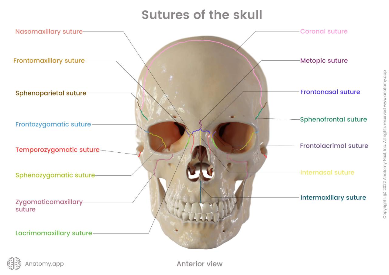
Sutures of the Skull Quiz: Dive into the fascinating world of human anatomy and test your understanding of the intricate sutures that hold our skulls together. From the coronal suture to the lambdoid suture, this quiz will challenge your knowledge and reveal the significance of these crucial connections.
Prepare to unravel the mysteries of the human skull, explore the functions of sutural bones and fontanelles, and discover the clinical importance of sutures in diagnosing and treating skull disorders. Let the quiz begin!
Sutures of the Skull: Sutures Of The Skull Quiz

The skull is a complex structure made up of several bones that are connected by joints called sutures. These sutures are important for allowing the skull to grow and change shape as we grow, and they also help to protect the brain from injury.
paragraphThere are several different types of sutures in the human skull, each with its own unique location and function. The most common type of suture is the serrated suture, which is found between the bones of the skullcap. Serrated sutures have a zigzag pattern that helps to interlock the bones and prevent them from moving.
Another type of suture is the squamous suture, which is found between the bones of the skull base. Squamous sutures are flat and smooth, and they allow the bones to slide against each other slightly.
Types of Sutures
- Serrated sutures: These sutures have a zigzag pattern that helps to interlock the bones and prevent them from moving.
- Squamous sutures: These sutures are flat and smooth, and they allow the bones to slide against each other slightly.
- Beveled sutures: These sutures have a slanted edge that helps to hold the bones in place.
- Butt sutures: These sutures are found between the bones of the skull base. They are flat and do not allow any movement between the bones.
Major Sutures of the Skull
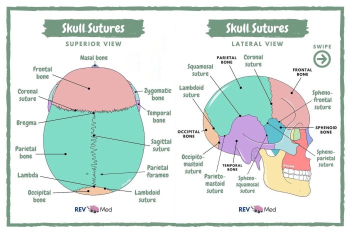
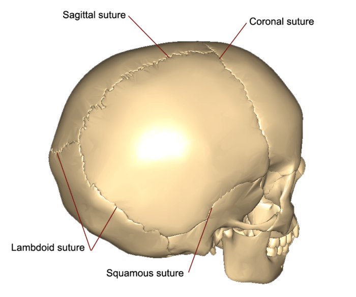
The skull is composed of multiple bones that are connected by fibrous joints called sutures. These sutures allow for some movement and growth of the skull during development. The major sutures of the skull include the coronal, sagittal, lambdoid, and squamous sutures.
Coronal Suture
The coronal suture is located at the junction of the frontal and parietal bones. It runs from one temporal bone to the other, across the top of the skull. The coronal suture is a serrated suture, which means that it has a zigzag shape.
This shape allows for some movement between the frontal and parietal bones, which is necessary for brain growth.
Sagittal Suture
The sagittal suture is located at the junction of the two parietal bones. It runs from the coronal suture to the lambdoid suture, along the midline of the skull. The sagittal suture is a straight suture, which means that it does not have a zigzag shape.
This suture is immovable, which helps to protect the brain from injury.
Lambdoid Suture
The lambdoid suture is located at the junction of the parietal and occipital bones. It runs from the mastoid process of the temporal bone to the occipital protuberance. The lambdoid suture is a serrated suture, which allows for some movement between the parietal and occipital bones.
This movement is necessary for brain growth and for the passage of the head through the birth canal.
Squamous Suture
The squamous suture is located at the junction of the temporal and parietal bones. It runs from the zygomatic process of the temporal bone to the mastoid process of the temporal bone. The squamous suture is a serrated suture, which allows for some movement between the temporal and parietal bones.
This movement is necessary for brain growth and for the passage of the head through the birth canal.
Sutural Bones and Fontanelles

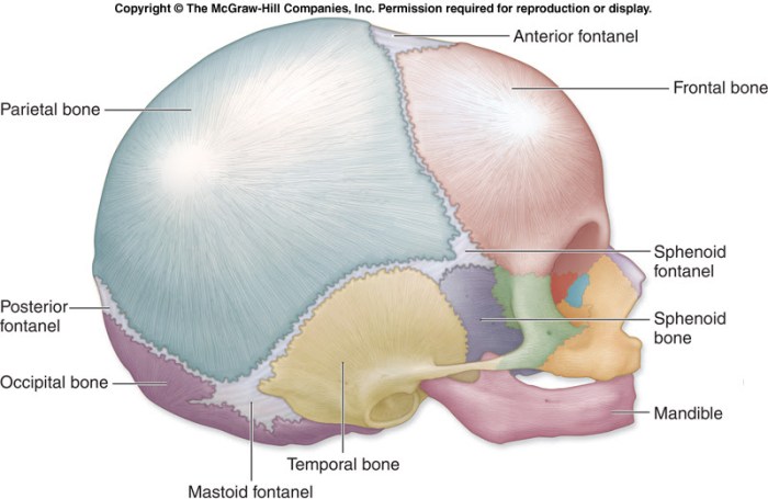
Sutural bones are small, irregular bones that form within the sutures of the skull. They play a crucial role in the development of the skull, providing flexibility and allowing for growth and expansion of the brain. The presence of these bones also helps to absorb impact and protect the brain from injury.Fontanelles
are soft, membranous areas on the skull of infants. They are located at the intersections of the skull sutures and allow for the skull to expand and accommodate the rapidly growing brain. The fontanelles typically close within the first two years of life as the skull fuses together.
Major Sutural Bones
* Inca bone: A small bone located at the intersection of the coronal and lambdoid sutures. It is named after the Incas, who believed it to be a sacred bone.
Epipteric bone
For a comprehensive review of the sutures of the skull, don’t miss the detailed answer key provided in the capitulo 3a 1 answer key . This resource will further enhance your understanding of the complex anatomy and functions of these cranial sutures.
A small bone located at the intersection of the parietal and temporal bones. It is often present in children and may persist into adulthood.
Significance of Fontanelles
Fontanelles are essential for the proper development of the brain. They allow for the skull to expand as the brain grows and provide a soft spot for the head to mold during birth. The closure of the fontanelles indicates that the skull has reached its adult size and the brain has stopped growing.
Clinical Significance of Sutures

Sutures play a crucial role in the diagnosis and treatment of various skull disorders. They provide essential clues about the growth and development of the skull and can indicate underlying medical conditions.
Role in Skull Growth and Development
Sutures are dynamic structures that allow for the growth and expansion of the skull during childhood. By monitoring the closure of sutures, clinicians can assess the normal development of the skull and identify any abnormalities that may indicate growth disorders.
Diagnosis of Skull Disorders
Abnormal suture closure can be a sign of underlying medical conditions, such as:
- Craniosynostosis: Premature closure of one or more sutures, leading to an abnormal shape of the skull.
- Plagiocephaly: Asymmetrical skull shape due to uneven growth of the skull bones.
- Microcephaly: Abnormally small head size due to premature closure of multiple sutures.
By examining the sutures and their closure patterns, clinicians can diagnose these disorders early on and initiate appropriate treatment.
Treatment of Skull Disorders
In some cases, surgical intervention may be necessary to correct abnormal suture closure and restore the normal shape of the skull. Surgery involves releasing the prematurely fused sutures to allow for proper growth and development.
Imaging of Sutures

Imaging techniques play a crucial role in visualizing and evaluating the sutures of the skull. These techniques provide valuable insights into the integrity, alignment, and development of the sutures, aiding in the diagnosis and management of skull disorders.
X-rays
- Advantages:
- Widely available and cost-effective
- Can detect gross abnormalities, such as suture widening or obliteration
- Limitations:
- Limited soft tissue contrast
- Can miss subtle suture abnormalities
CT scans
- Advantages:
- Excellent bone detail
- Can detect subtle suture abnormalities, such as synostosis or dehiscence
- Can visualize adjacent structures, such as the brain and sinuses
- Limitations:
- Higher radiation exposure than X-rays
- May not be suitable for infants or young children
MRIs
- Advantages:
- Excellent soft tissue contrast
- Can visualize suture patency and detect subtle abnormalities
- Can assess the surrounding brain and other structures
- Limitations:
- More expensive and time-consuming than other imaging modalities
- May not be suitable for patients with metal implants or claustrophobia
Example: Diagnosis of Skull Disorders, Sutures of the skull quiz
Imaging findings can aid in the diagnosis of various skull disorders, such as:
- Craniosynostosis: Premature fusion of one or more sutures, leading to an abnormal skull shape
- Craniolacunia: A condition where sutures fail to fuse properly, resulting in gaps in the skull
- Suture diastasis: A widening of a suture, often associated with increased intracranial pressure
FAQ Guide
What is the function of sutures in the skull?
Sutures allow for slight movement and growth of the skull, especially during infancy, and provide a pathway for blood vessels and nerves to pass through.
What is the largest suture in the skull?
The sagittal suture, located along the midline of the skull, is the longest and most prominent suture.
What is the significance of fontanelles in infants?
Fontanelles are soft spots in the infant skull that allow for brain growth and the molding of the skull during birth.