Exercise 29 review sheet anatomy of the urinary system – Embarking on a journey to unravel the intricate workings of the urinary system, this review sheet provides a comprehensive guide to its anatomy. From the kidneys’ role in waste filtration to the intricate network of ureters, bladder, and urethra, this exploration delves into the structural foundations of this vital bodily system.
The urinary system, a complex machinery of organs, plays a pivotal role in maintaining homeostasis and eliminating waste products from the body. Understanding its intricate anatomy is essential for healthcare professionals and medical students alike, as it serves as the cornerstone for diagnosing and treating urinary tract ailments.
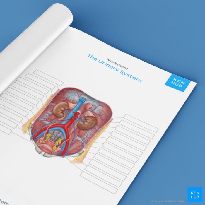
Overview of the Urinary System

The urinary system plays a crucial role in maintaining the body’s fluid and electrolyte balance, regulating blood pressure, and removing waste products. It consists of two kidneys, two ureters, a urinary bladder, and a urethra.
- Kidneys:Filter waste products from the blood and produce urine.
- Ureters:Transport urine from the kidneys to the bladder.
- Urinary Bladder:Stores urine until it is released during urination.
- Urethra:Conducts urine from the bladder to the outside of the body.
Anatomy of the Kidneys, Exercise 29 review sheet anatomy of the urinary system
External Anatomy:Bean-shaped organs located on either side of the spine. The outer layer, called the renal capsule, protects the kidneys.
Internal Anatomy:The kidneys are divided into an outer cortex and an inner medulla. The cortex contains the nephrons, the functional units of the kidneys. The medulla contains the collecting ducts, which collect urine from the nephrons.
Nephrons:Microscopic structures responsible for filtering waste products from the blood. Each nephron consists of a glomerulus, a Bowman’s capsule, and a renal tubule.
Urine Formation:Urine is formed through a process called glomerular filtration, tubular reabsorption, and tubular secretion. Blood is filtered through the glomerulus into the Bowman’s capsule. Essential substances are reabsorbed back into the blood in the renal tubule, while waste products are secreted into the tubule.
The resulting fluid is urine.
Ureters, Urinary Bladder, and Urethra
Ureters:Narrow tubes that transport urine from the kidneys to the bladder. They are lined with smooth muscle, which allows them to contract and push urine downward.
Urinary Bladder:A muscular sac that stores urine until it is released during urination. The bladder wall contains a layer of smooth muscle called the detrusor muscle, which contracts to empty the bladder.
Urethra:A short tube that carries urine from the bladder to the outside of the body. In males, the urethra is also part of the reproductive system and transports semen during ejaculation.
Microscopic Anatomy of the Urinary System
Kidneys:The renal cortex contains glomeruli, Bowman’s capsules, and proximal and distal convoluted tubules. The renal medulla contains collecting ducts and loops of Henle.
Ureters, Urinary Bladder, and Urethra:The walls of these structures are lined with transitional epithelium, which can stretch to accommodate changes in volume.
Relationship to Function:The microscopic anatomy of the urinary system reflects its function. The glomeruli are responsible for filtering waste products from the blood, while the tubules reabsorb essential substances and secrete waste products. The smooth muscle in the ureters and bladder allows for the transport and storage of urine.
Clinical Significance of Urinary System Anatomy
Understanding the anatomy of the urinary system is crucial for diagnosing and treating urinary tract infections, kidney stones, and other disorders.
- Urinary Tract Infections:Infections of the urethra, bladder, or kidneys. Understanding the anatomy of these structures helps in identifying the source of the infection and guiding treatment.
- Kidney Stones:Hard deposits that form in the kidneys. Knowledge of kidney anatomy aids in localizing the stones and planning their removal.
- Imaging Techniques:X-rays, ultrasound, and CT scans are used to visualize the urinary system. Understanding the anatomy helps in interpreting these images and identifying abnormalities.
Question Bank: Exercise 29 Review Sheet Anatomy Of The Urinary System
What are the primary functions of the urinary system?
The urinary system is responsible for filtering waste products from the blood, regulating fluid balance, and maintaining electrolyte balance.
Describe the structure and function of the nephrons.
Nephrons are the functional units of the kidneys. They filter waste products from the blood and produce urine.
Explain the role of the urinary bladder in urine storage.
The urinary bladder is a muscular organ that stores urine until it is released through the urethra.